Services Case Studies State Of The Art Facility  

Acoustic Neuroma

A gamma knife case study: Acoustic Neuroma

Radiation Oncology 2011

Patient Presentation

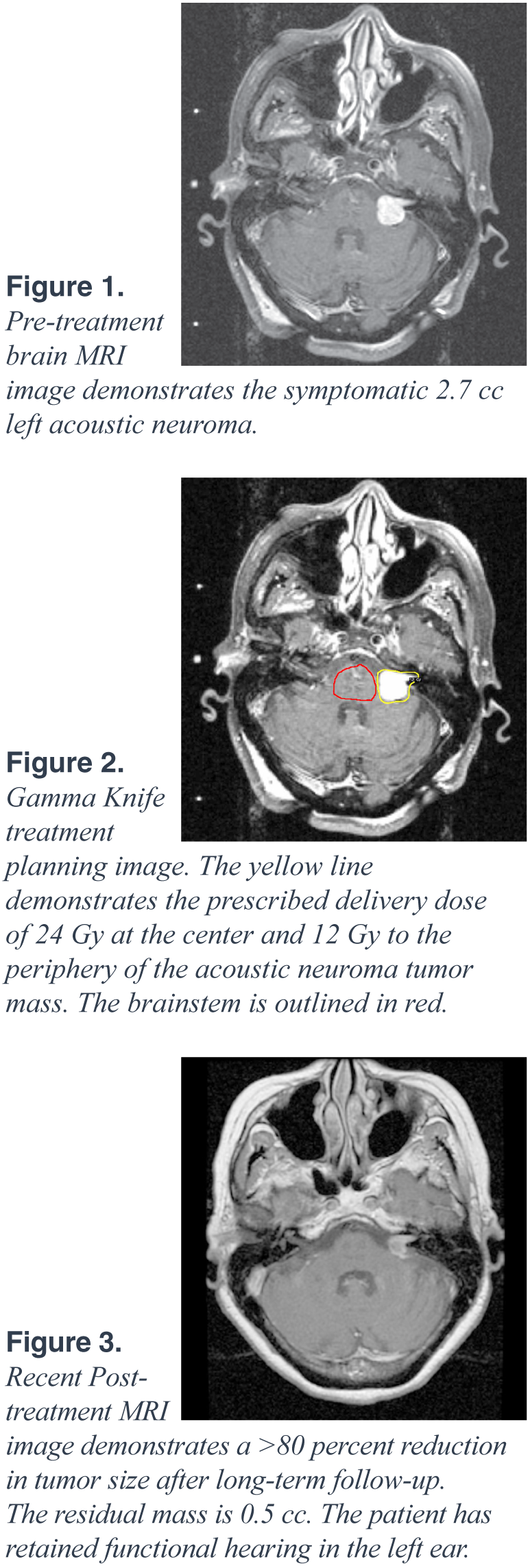
The patient is a 75-year-old female who presented to Neurosurgeon Dr. Asher Taban, MD, and Radiation Oncologist Afshin Safa, MD, with a five-year history of left-sided tinnitus and progressive sensorineural hearing loss. An MRI of the brain was performed, which revealed a 2.7 cc left-sided enhancing mass compressing the brainstem and extending from the left internal auditory canal into the cerebellopontine angle region. The radiographic features of this mass were consistent with an acoustic neuroma, which is a common benign tumor of the eighth cranial nerve. Her case was presented to our multidisciplinary Gamma Knife Review Board. Given the tumor’s size, location, and the associated symptoms of hearing loss and tinnitus, the patient underwent Gamma Knife Stereotactic Radiosurgery at Northridge Hospital Medical Center.

Procedure

On the day of treatment at the Gamma Knife Center, a stereotactic head frame was fitted and a treatment planning MRI was obtained. A three-dimensional treatment plan was developed on the Gamma Knife treatment planning workstation to deliver an ablative dose of 24 Gy at the center and 12 Gy to the periphery of the tumor, while sparing the adjacent brainstem. The radiation dose was delivered using 15 discrete spherical shots to sculpt the dose to conform to the acoustic neuroma and avoid the adjacent normal structures. The Radiosurgery treatment was delivered in less than one hour. The patient tolerated the procedure well and was able to return to normal activities later that day.

Follow-up

We have continued to follow the patient on a semiannual basis since her Gamma Knife Stereotactic Radiosurgery. She has had improvement of her tinnitus and has retained functional hearing in the left ear. Three-years after treatment, her most recent MRI revealed a 0.5 cc residual mass with necrotic center, which represents a greater than 80 percent reduction in size, as well as diminished radiographic enhancement. The patient maintains her active life and is a world traveler. She will continue to be followed by our Gamma Knife treatment team on a semiannual basis.


Gamma Knife Radiosurgery

Amazing Recovery and Survival with Gamma Knife Radiosurgery

Neuroscience Innovations, Vol. 5, 2010

Patient Presentation

A patient with colon cancer and multiple metastatic brain tumors has survived since 2005 after Gamma Knife Radiosurgery. This is just one of our amazing success stories. Brain metastasis from colorectal cancer is uncommon with incidence of 1.5 – 2.3 percent of all brain metastasis. The cerebellum is the most common area of colon cancer to brain metastasis and left-sided primary colon tumors predominate. Increased survival among patients with metastatic colorectal carcinoma will likely result in an increased incidence of brain metastases.

Procedure

Whole brain radiotherapy survival ranges from 3–6 months. However, Gamma Knife Radiosurgery may result in a much higher survival rate and less brain toxicity. We treated such a patient at Northridge Hospital in 2005, who has remained fully functional with no identifiable brain lesion to date. This 77-year-old white male patient was first diagnosed with colon cancer in 1993 and treated with surgery and chemotherapy. He did well until early 2005 when he was found to have two enhancing brain lesions, including the left cerebellar tonsil measuring 21x19x16 mm and right parietal lobe measuring 11x12x14 mm with surrounding edema. The patient underwent Gamma Knife Radiosurgery at Northridge Hospital on June 30, 2005 and continues to remain under close observation.

Follow-up

The most recent MRI scan of the brain on November 12, 2009 revealed no identifiable metastatic tumor with 6–7 mm area of gliosis in the left inferior cerebellum, unchanged in the past four years. The original mass in this region measured 21mm in diameter. The patient is fully functional and remains neurologically intact. Factors influencing survival include Karnofsky performance scale score over 70, status of systemic disease and total number of intracranial tumors and histological diagnosis. The dramatic survival of this patient is an example of the efficacy of Gamma Knife Radiosurgery versus whole brain radiation.


© 2013 Asher Taban, MD Disclaimer | Website Design by Infinite Communications